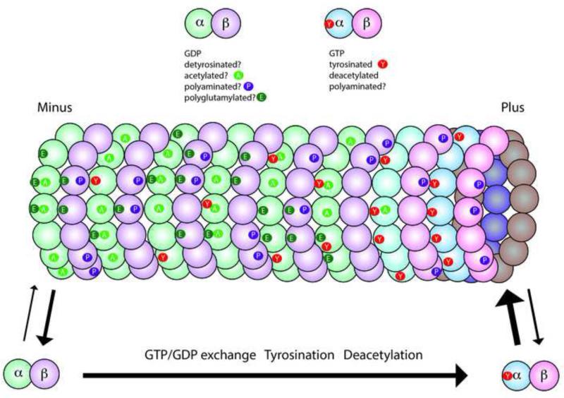
Figure 1. Microtubules and tubulins are subject to a variety of posttranslational modifications.
This diagram illustrates the major modifications in axonal microtubules. Tubulin dimers are GTP-rich (blue/pink shading) in the soluble pool and on the plus end of the microtubule, but polymeric tubulin gradually hydrolyzes GTP and becomes GDP-rich (green/purple shading). Some modifications are associated specifically with tubulin polymerized in microtubules including acetylation (A), detyrosination, and polyglutamylation (E), while others may occur only on soluble tubulin like tyrosination (Y), or on either soluble or polymerized tubulin like polyamination (P). Some tubulins will have a single modification, while others have multiple modifications and some have no modification. However, the exact distribution along a microtubule and the fraction of the tubulin modified is highly variable, even within a single cell. A few rules have emerged such as tyrosinated tubulin is enriched at the plus ends of growing microtubules, while acetylation and detyrosination are more likely to be seen in the middle or minus end of the microtubule.

