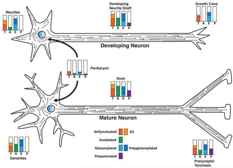
Figure 5. Posttranslational modifications of tubulin and microtubules vary in different regions of a neuron and change during neuronal differentiation.
Microtubules in neuronal axons and dendrites are not anchored at the microtubule organizing center. The microtubules vary in length but have both plus and minus ends free. In the developing neuron (upper), when the axon is extending but the dendrites have not yet differentiated, the microtubules in all neurites are plus end out. Modification levels for detyrosination (T), Δ2 tubulin (Δ2), glutamylation (E), and polyamination (P) in the neuronal perikaryon are relatively low. Acetylation, glutamylation and detyrosination are elevated in the growing axon, but all three are reduced in the growth cone. Little or no acetylation is seen in the microtubules of the growth cone, consistent with the presence of highly dynamic microtubules. The minor neurites at this stage are lower in acetylation, but relatively rich in detyrosinated and glutamylated/polyglutamylated microtubules. Polyamination may be detectable in the growing axon at a low level.
In a mature neuron (lower), as both dendrites and synaptic specializations form, tubulin modifications change both quantitatively and qualitatively. Dendritic microtubules exhibit mixed polarity as dendrites form, while axonal microtubules remain plus end out. Polyamination of axonal microtubules increases with maturation, but may be relatively low or absent from dendritic domains. Detyrosinated and Δ2 tubulin levels remain relatively high in differentiated dendrites and axons, while acetylation increases in both axon shafts and dendrites. Acetylation is also detectable in the distal axon and presynaptic regions, consistent with reduced numbers of highly dynamic microtubules in stable connections. During the differentiation and maturation of neurons, there are also changes in microtubule associated proteins in different neuronal subcellular domains. Adapted from Janke and Kneussel [5].

