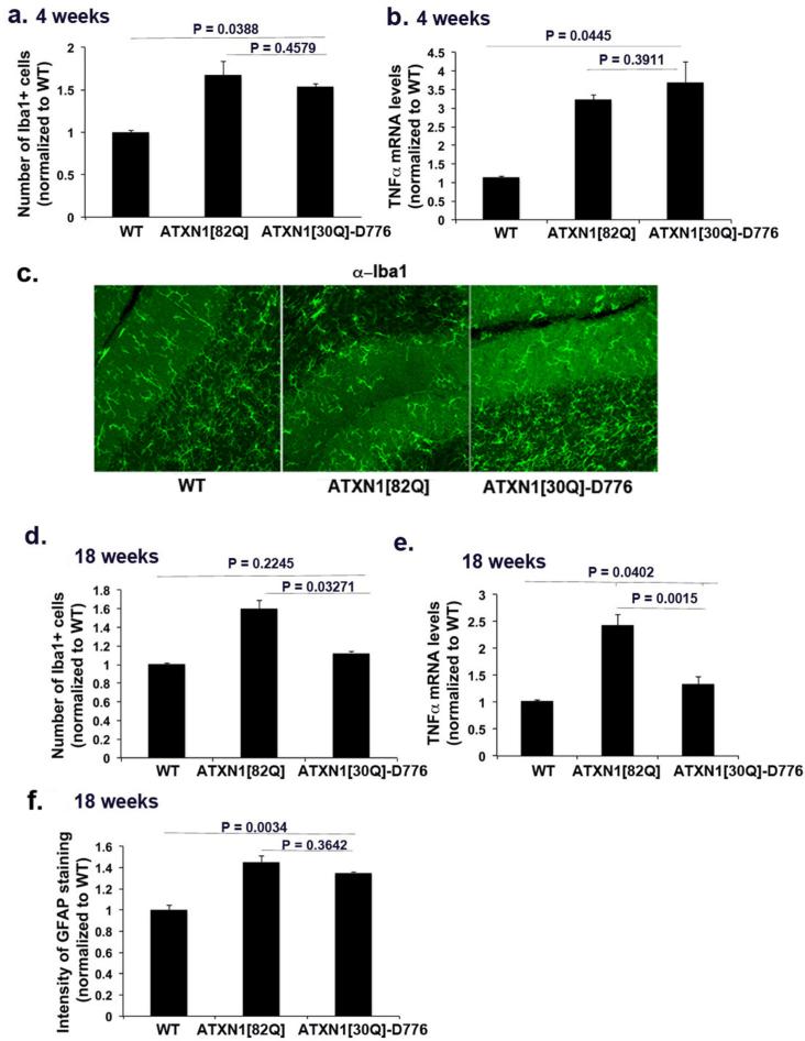
Figure 5. Microglial activation correlates with loss of Purkinje neurons in SCA1.
Cerebellar sections from 4 or 18 weeks old wild-type (WT), ATXN1[82Q], and ATXN1[30Q]-D776 mice were stained for Iba1 (c representative image) and GFAP. a and d Number of Iba1+ microglia in cerebellar cortex at 4 weeks (a) and 18 weeks (d). f GFAP intensity at 18 weeks. b and e Quantitative RT qPCR was used to determine relative levels of TNFα in 4 (b) or 18 weeks old (e) wild-type (WT), ATXN1[82Q], and ATXN1[30Q]-D776 cerebella. N ≥ 5 mice were used for all experiments. Student's t-test P values are shown with all the histograms. Error bars represent SEM.

