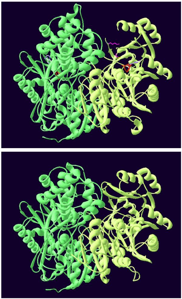
Figure 9.
Modelled structure of the S. cerevisiae CPSase domain of the CPSase-ATCase complex. Top: cristallographic structure of the E. coli CPSase [32]. Bottom: modelled structure of the S. cerevisiae CPSase domain of the bifunctional protein obtained as indicated in the Methods. The CPS-A and CPS-B domains are in green and light-green respectively. The two catalytic subsites of the E. coli CPSase are indicated by two molecules of ADP. Lysine992 and serine948 located in the UMP binding site of the E. coli CPSase are in magenta.

