
Figure 1. Topological Model of MHC-II movement from intralumenal vesicles of MVB to the plasma membrane.
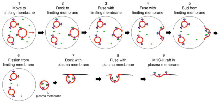
MHC-II are present on lipid raft intraluminal vesicles (ILVs) with their peptide-binding grooves exposed to the antigenic peptide-containing lumen of the MVB. A pMHC-II containing ILV (indicated by red membrane) moves toward the MVB limiting membrane (step 1), docks with (step 2) and fuses with the limiting membrane (steps 3 & 4). Continued outward budding from the limiting membrane (step 5) leads to ILV fission from the limiting membrane and release of a membrane-derived transport vesicle (step 6). This transport vesicles docks with the plasma membrane (step 7) and the docked membranes fuse (steps 8 & 9), thereby depositing a “MHC-II raft” generated from ILV membrane into the plasma membrane.