
Figure 3. Newly-arrived pMHC-II complexes are clustered on the DC surface.
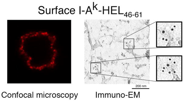
Immature DCs were incubated in with HEL protein, washed, and activated with LPS for 12 hr. The DCs were stained live with a mAb that only recognizes the I-Ak-HEL(46–61) complexes. Aw3.18.14. The cells were analyzed by confocal immunofluorescence microscopy and a single 0.8 μm thick optical section is shown (left panel). The distribution of these newly-arrived pMHC-II complexes on the DC surface was also determined using “plasma membrane rips” and analysis by immunoelectron microscopy (right panel) (Reprinted with permission from reference [16].