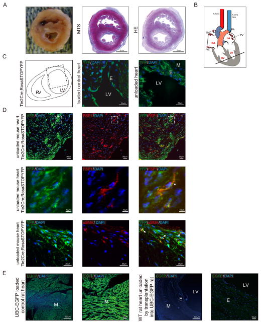
Figure 4. EFE in the Tie2Cre;Rosa-Stop-YFP reporter mouse is derived from EndMT.
(A) Representative macroscopic picture and photomicrographs of Masson’s Trichome and HE stained sections of unloaded rodent hearts after heterotopic transplantation. (B) Sketch of the rodent unloaded heterotopic heart transplant model, the bold line indicates the position of the cross-sections (C) Sketch of heart cross-section, dotted line rectangle indicates the area used for confocal microscopy (left panel). Representative confocal photomicrographs of heart sections from neonatal unloaded Tie2Cre; Rosa-Stop-YFP mice two weeks after heterotopic transplantation (right panel) and Tie2Cre; Rosa-Stop-YFP control hearts (middle panel). (D) Representative confocal photomicrographs of sections from neonatal unloaded heterotopic transplanted Tie2Cre; Rosa-Stop-YFP mouse heart stained for mesenchymal marker FSP1 (red, upper panel) or αSMA (red, bottom panel). Endogenous YFP expression is shown in green. Dotted areas denote the region which is shown magnified in the middle panel; white arrows denote representative double positive cells. (E) Representative confocal photomicrographs of sections from loaded control UBC-EGFP rat heart and unloaded rat heart. M, myocardium; E, endocardium; LV, left ventricular lumen.

