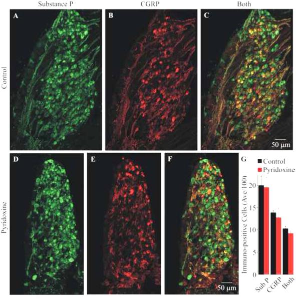
Fig. 2.
Effect of pyridoxine treatment on Substance P and CGRP immuno-reactivity in the third lumbosacral DRG. Panels (A-C) show images of the immuno-reactivity for Substance P (green), CGRP (red) and a superposition of the images, respectively, from the medial section of a control DRG. Panels (D-F) show similar images from the medial section of a pyridoxine-treated embryo. All images are maximum projections from optical sections collected using structured illumination. Notice that the treated DRG is smaller than the control DRG. The total number of cells (uc) from all embryos (mean and standard deviation) expressing Substance P, CGRP or both are shown in (G). There was no significant loss of Substance P or CGRP expressing cells.

