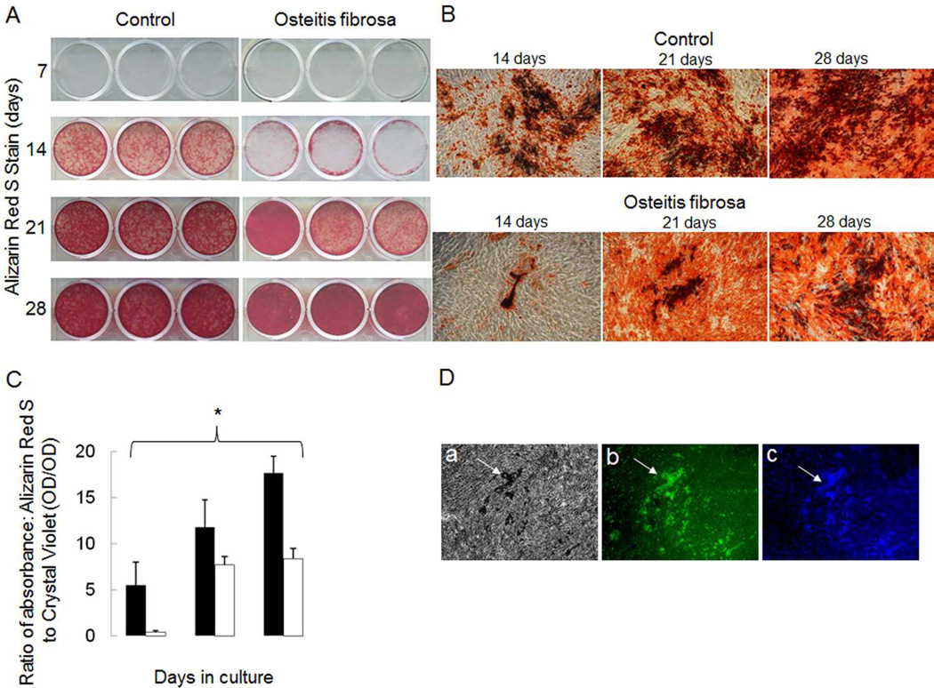
Figure 5.
Mineralization rates in cells from dialysis patients with high turnover renal osteodystrophy as compared to those from healthy controls. A) Alizarin Red S staining of primary human osteoblast-like cells from healthy controls and dialysis patients with high turnover renal osteodystrophy grown under mineralizing conditions (10 mM β-glycerolphosphate and 100 µg/ml ascorbic acid) for 7, 14, 21 and 28 days. B) Alizarin red stained mineralized nodules under light microscopy (40x) at 14, 21, and 28 days. C) Mineralization of primary osteoblast-like cells, as quantified by the ratio of absorbance of Alizarin Red S to Crystal violet (i.e. ratio of mineral content to cells), occurs more rapidly in cells from healthy controls (black bars) than in dialysis patients with high turnover renal osteodystrophy (open bars). The asterisk denotes significant (p<0.01) difference in Alizarin Red S to Crystal Violet absorbance ratios between dialysis patients with high turnover renal osteodystrophy and healthy controls. D) Primary osteoblast-like cells from healthy controls after 28 days in mineralizing conditions a) display mineralized nodules under light microscopy and b) express osteocalcin (green color) as detected by immunofluorescence. DAPI staining (c) confirms that osteocalcin expression localizes to living cells at the site of the mineralized nodule formation.

