
Figure 3.
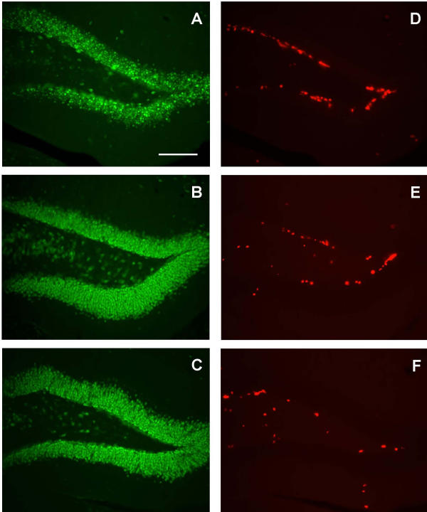
BrdU labeled cells in the dentate gyrus of the hippocampus. Photomicrographs showing representative NeuN staining of the dentate gyrus in a wild-type (A), a vehicle-treated R6/2 (B) and an asialoEPO-treated R6/2 (C) mouse of 12 weeks of age. Photomicrographs showing representative BrdU labeling in adjacent sections of the dentate gyrus in a wild-type (D), a vehicle-treated R6/2 (E) and an asialoEPO-treated R6/2 (F) mouse. Scale bar 100 μm.