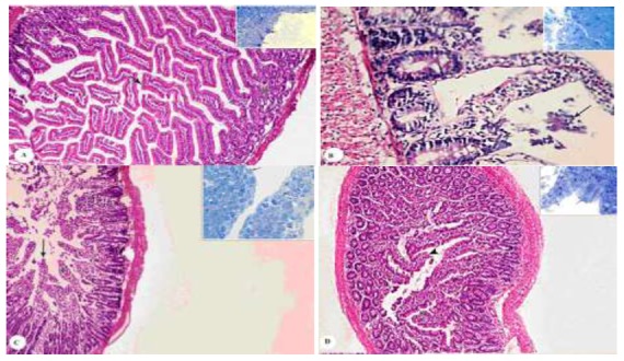
Fig. 1.

Sections in the small intestinal mucosa of control and treated rat groups. (A) From GI showing upright villi (arrow) and invaginated crypts (astrik). (H&E. X 100). Inset of a semithin section in the same group showing regular continuous brush border with tall columnar enterocytes (arrow) and goblet cells in-between (two arrows) (T.B (Toludine Blue) X1000). (B) From GII showing disorganized villi with pyknotic villous membrane and necrosis of some enterocytes with pyknotic nuclei (arrow), heavily infiltrated lamina propria with aggregated lymphocytes (astrik) (H&E. X 1000). Inset a semithin section in the same group showing villi fusion, blunting, erosion and adhesion (arrow), goblet cells can be seen (astrik) (T.B X1000). (C) From G IV after Ginger (20 mg/kg/day) showing few short columnar enterocytes covering areas of the villi (arrow), decrease in the inflammatory infiltration in the lamina propria (astrik) (H&E. X 100). Inset of a semithin section in the same group showing some cuboidal cells lining the villi (arrow), with moderate inflammatory cells (astrik) (T.B X1000). (D) From G VI after Cinnamon (20 mg/kg/day) showing normal tall, finger-like villi, most of them were covered with tall columnar enterocytes and goblet cells (arrow), but some villi have dislodged tips (arrow head) (H&E. X 100). Inset of a semithin section in the same group showing regular continuous brush border (arrow), other areas showed flat surface epithelial cell of the villi (arrow head) with mild inflammatory cells infiltrated the lamina propria (astrik) (T.B X1000)
