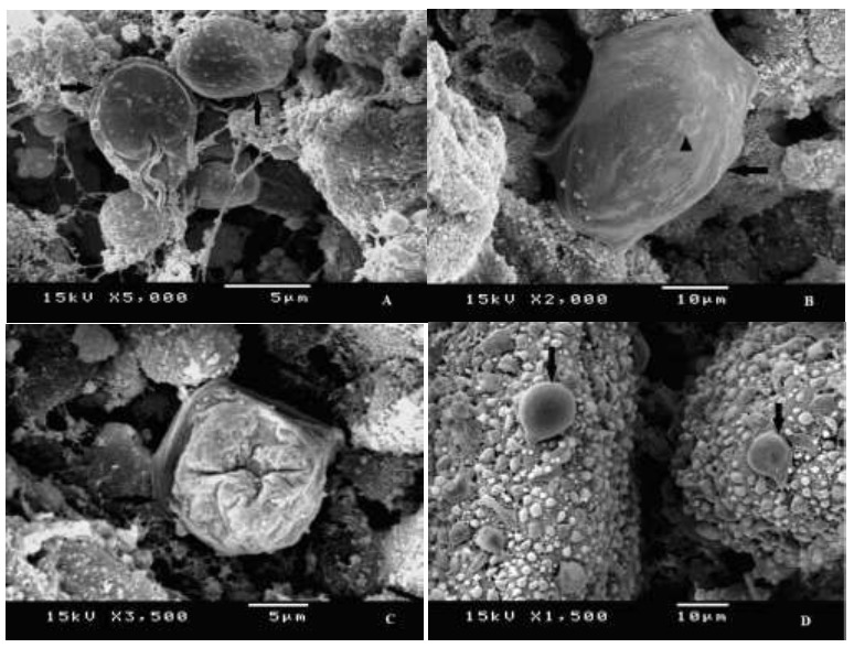
Fig. 3.

SEM of G. lamblia trophozoite (A) From GII (infected non-treated) showing normal trophozoites with smooth intact ventral and dorsal surface (arrows). (B) From G III (after Ginger 10 mg/kg/day) showing swollen trophozoite, still attached in situ (arrow) with multiple erosions (arrow head). (C) From G III (after Ginger 20 mg/kg/day) showing irregularities of the trophozoite, with complete peeling of the outer surface (arrow). (D) From G VI (after Cinnamon 20 mg/kg/day) showing swollen pear-shaped trophozoites (arrows)
