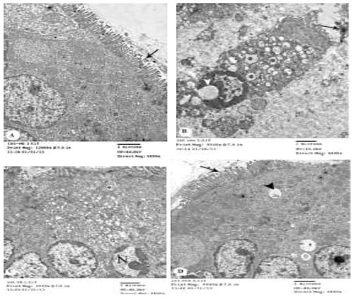
Fig. 4.

TEM of small intestinal mucosa of control and treated rat groups. (A) From GI (non-infected non-treated) showing typical appearance of microvillus brush border (arrow) with numerous mitochondria (astrik) (X5800). (B) From GII (infected non-treated) showing the separate laminated vesicle (arrow), the disruption of nuclei (arrow head) with increased peripheral chromatin. Notice the mitochondrial cristiolysis and loss of mitochondrial dense matrix in the supranuclear region (astrik) (X 4800). (C) From GIV after (Ginger 20 mg/kg/day) showing enterocytes with membrane-bound cytoplasmic vacuoles (astrik). Notice the disruption and polymorphism of nuclei (N) with increased peripheral chromatin (X4800). (D) From G VI (Cinnamon 20mg/kg/day) showing the microvilli restored normal length, thickness, parallel orientation but irregular in height (arrow). The enterocytes regained normal mitochondrial (astrik) and nuclear chromatin density and electron-lucent Golgi vesicles reappeared near the lateral cell membranes. Notice the membrane-bound cytoplasmic vacuoles (arrow head) (X4800)
