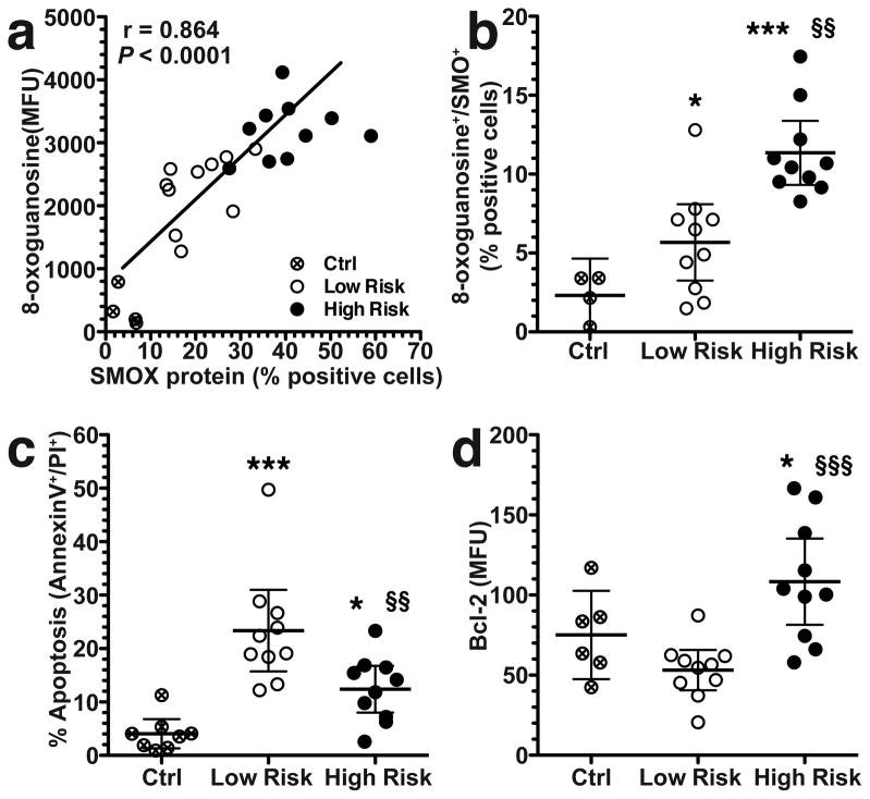
Figure 2.
Correlation between SMOX and DNA damage, and levels of apoptosis and Bcl-2 in AGS cells co-cultured with clinical isolates for 24 h. (a) Correlation (Pearson coefficient) between the percentage of cells positive for SMOX and 8-oxoguanosine levels determined by flow cytometry. (b) Percentage of SMOX+, 8-oxoguanosine+ double-positive cells by flow cytometry. (c) Summary data for apoptosis measured by annexin V and propidium iodide (PI) using flow cytometry. (d) Summary data for levels of Bcl-2 measured by flow cytometry. For ad, at least three experiments were performed in duplicate. In b–d, *P < 0.05, ***P < 0.001 versus control (Ctrl); §§P < 0.01, §§§P < 0.001 versus low risk. Ten strains from each risk group were used.

