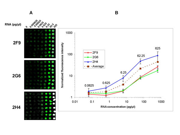
Figure 3.

Evaluation of dynamic range and sensitivity of RNA Arrays. Images from RNA arrays prepared from 10-fold dilution series of three external control RNAs (2F9, 2G6 and 2H4) and hybridized with Cy3 labeled probes (A). Each of the three plant RNAs were spiked into rat liver total RNA (0.5 μg/μl) with 10 fold dilution series ranging from 0.000625 – 625 pg/μl. The first column is negative control (liver total RNA with no spiked in RNA). Log-log plots of normalized average fluorescence intensity of 8 replicate spots per dilution versus RNA concentration in the dilution series of the three transcripts (B). The two lowest concentrations 0.000625 and 0.00625 pg/μl did not result in signals over background levels and were omitted in the plots. Data are presented as mean ± SD.
