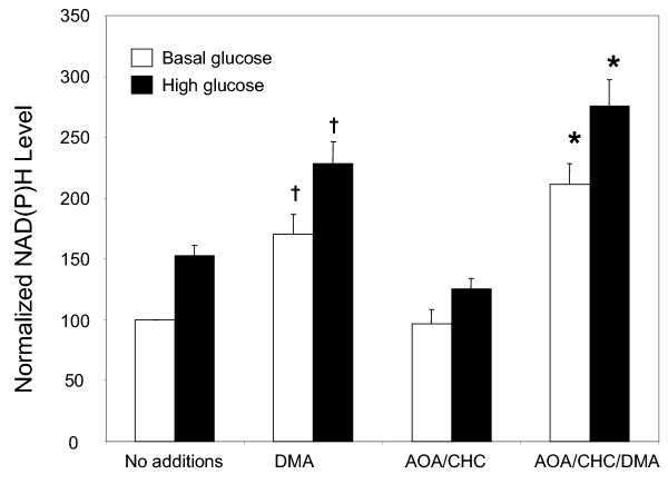
Figure 7.
DMA increases whole cell NAD(P)H, both in the presence and absence of inhibitors: Islets were placed in basal KRBH with or without mitochondrial inhibitors (AOA and CHC, 5 mM each) and/or 40 μM DMA, and NAD(P)H auto-fluorescence was measured by TPEM before and after addition of 16.7 mM glucose. NAD(P)H auto-fluorescence for each condition is expressed as a percentage of the control (basal glucose with no additions). n = 6; † = p < 0.05 and * = p < 0.002, when compared to the same condition without DMA.

