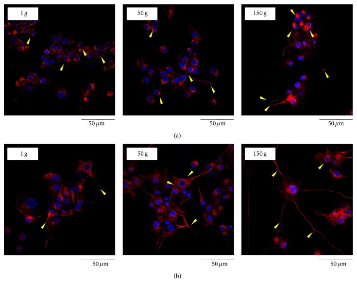
Figure 6.
Confocal images of neurofilament-66 (in red) and nuclei (in blue) in PC12 cell cultures predifferentiated for 12 h (a) and 72 h (b) after 48 h from hypergravity treatment. Arrows evidence neurofilament-66 organization in a ring-like structure in control cultures, whereas the marker is localized in neurites and in growth cones in hypergravity-stimulated cells.

