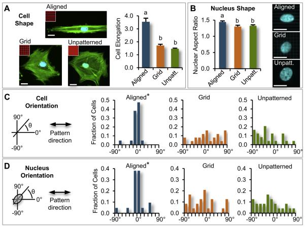
Fig. 1.
Micro-photopattern (μPP) micro-scale architecture directs alignment and shape of single hMSCs. (A) hMSCs were seeded on μPP substrates with aligned, grid, or unpatterned micro-scale cell-adhesive architectures (green:actin; blue:cell nuclei; red(inset):fluorescently-labeled fibronectin showing μPP architecture), with cell elongation (max/min Feret diameter) and cell nuclear aspect ratios measured following 2h attachment. Single hMSCs (not in contact with other cells) on aligned architectures were (B) more elongated (ANOVA, p < 0.0001, n = 21–25 cells/substrate condition, conditions labeled with different letters were statistically different) and (C) exhibited higher nuclear aspect ratios (ANOVA, p < 0.01) compared to cells on grid or unpatterned μPP architectures (representative images of cell nuclei shown on right). On aligned μPP architecture, cells (D) and cell nuclei (E) were significantly more aligned with pattern direction, in contrast to on grid or unpatterned architectures (*p < 0.01 compared to grid, unpatterned, Kolmogorov–Smirnov test). Scale bars = 20 μm. (For interpretation of the references to colour in this figure legend, the reader is referred to the web version of this article.)

