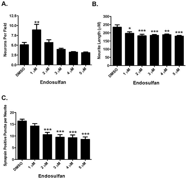
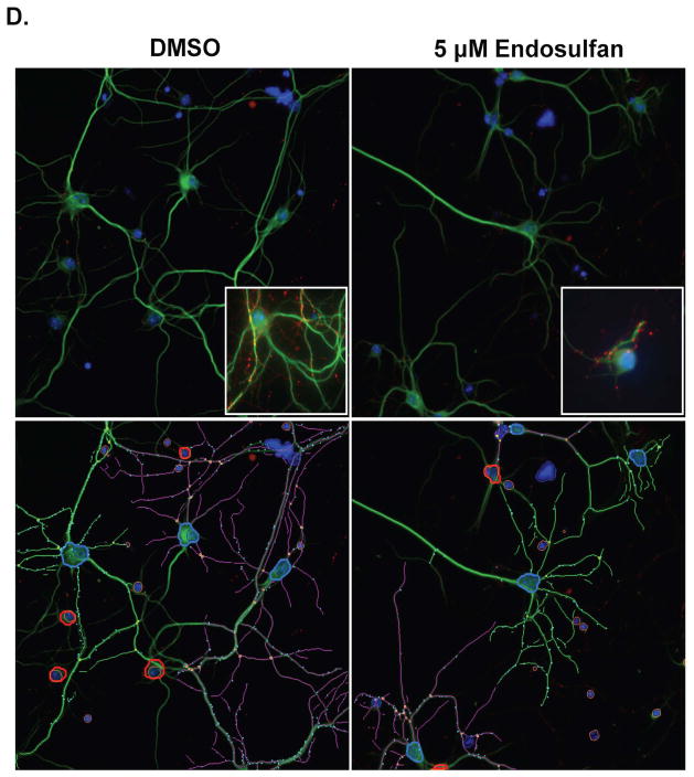
Figure 2.
Exposure of cortical primary cultures to endosulfan causes a loss of cortical neurons as well as reductions in neurite outgrowth and synapse formation. (A) Treatment of cortical cultures caused a significant reduction in the number of MAP2+ neurons, compared with DMSO control. (B) Assessment of neurite length in these neurons demonstrated a greater loss of neurite outgrowth compared with DMSO control. (C) Evaluation of synapse formation also showed a substantial reduction, compared with DMSO control. Columns represent the percent change from DMSO control for each concentration of endosulfan. (D) Representative images of primary cultured cortical neurons treated with DMSO or 5 μM endosulfan for 9 days and immunolabeled with MAP2 (green), synapsin (red), and Hoechst nuclear counterstain (blue). Insets represent higher magnification images of cortical cultures treated with DMSO or endosulfan. Images below are traces of the same images to highlight our use of the automated quantification of neuron number, neurite length, and the number of synapsin positve puncta in our DMSO and endosulfan treated cultures. Blue outline denotes a cell body that was selected for analysis. In contrast, red outline denotes cell bodies that did not meet our analysis parameters and were not selected or included for analysis. Green and purple traces represent neurites assigned to different cell bodies. Cyan dots denote synapsin positive puncta that were adherent to our parameters for analysis. Data represent the mean ± SEM of 17 experimental replicates per treatment group performed across 3 separate experiments. *Values for treatments significantly different from DMSO control (p < 0.05). **Values for treatments significantly different from DMSO control (p < 0.01). ***Values significantly different from DMSO control (p < 0.001).


