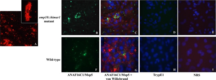
FIG 4.

Infection of RF/6A endothelial cells at 10 days postinfection with the omp10::himar1 mutant and wild-type Anaplasma marginale strains. (A) RF/6A endothelial cells infected with the A. marginale omp10::himar1 mutant expressing mCherry red fluorescent protein, with the inset showing a single endothelial cell infected with multiple colonies. The A. marginale (green) omp10::himar1 mutant (B and C) and the wild type (F and G) in endothelial cells (red). In panels C and G, host cell nuclei were counterstained with DAPI (blue). Similar fluorescence was not detected in infected cells stained with control antibodies Tryp1E1 or NRS for cells infected with the omp10::himar1 mutant (D and E) or in cells infected with the wild type (H and I).
