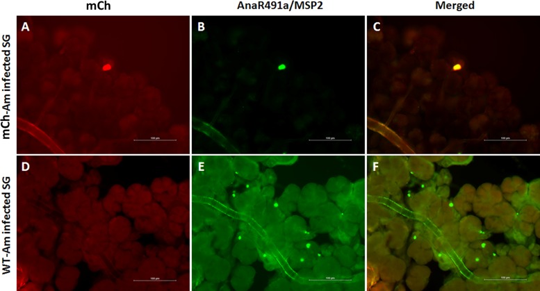
FIG 6.
The A. marginale omp10::himar1 mutant infects ticks. Immunofluorescence images showing infection of D. andersoni salivary gland (SG) acinar cells with the omp10::himar1 mutant (mCh-Am) (top) and wild-type A. marginale (WT-Am) control (bottom). (A to C) Salivary glands from tick acquisition fed on a calf infected with mCh-Am (red) were colabeled with AnaR4901a primary antibodies against Anaplasma marginale MSP2 and a conjugated Alexa Fluor 488 goat anti-rabbit IgG-H&L secondary antibody (green). (D to F) Salivary glands from control ticks infected with wild-type A. marginale showing multiple colonies of mCherry-negative, MSP2-positive A. marginale.

