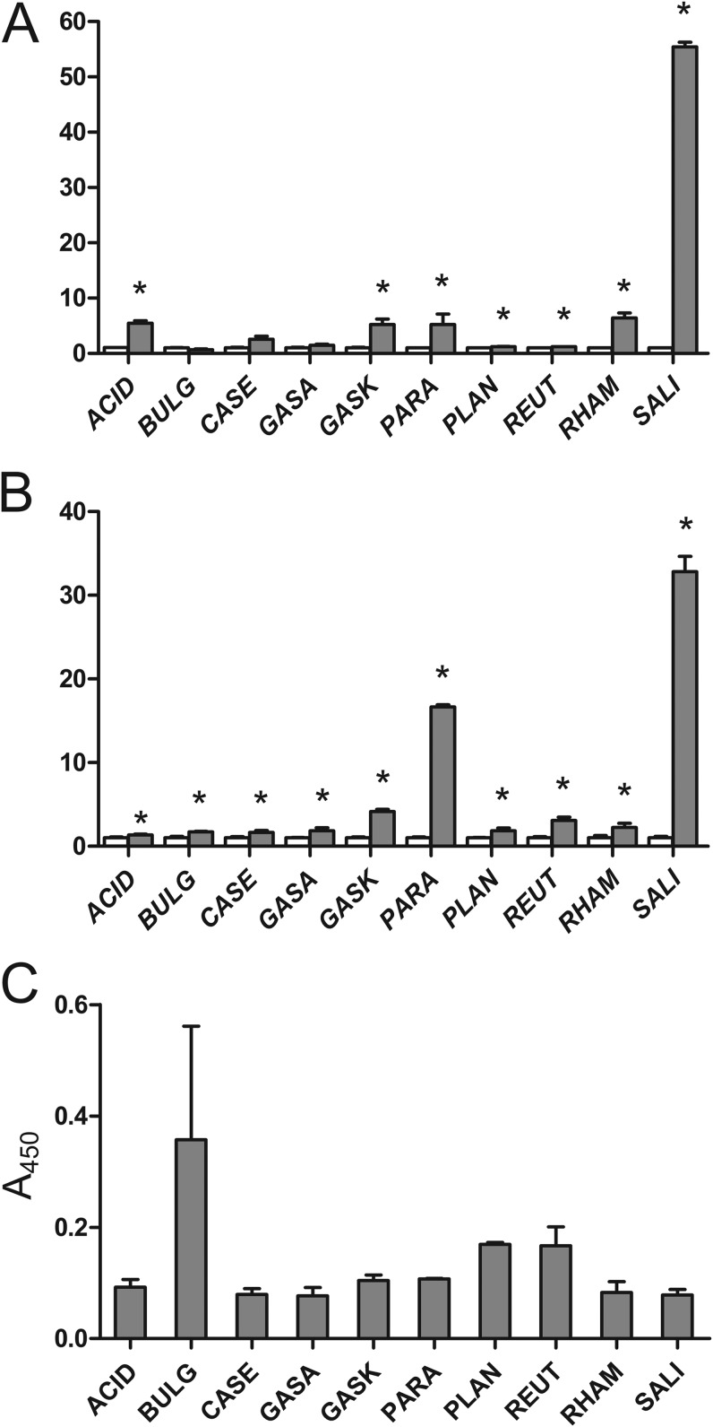
FIG 3.
Flow cytometric (A) and whole-cell ELISA (B) analyses of Lactobacillus sp. cells after incubation with DARPin I_07-cA fusion-containing growth medium and detection with FITC-conjugated human IgG (gray bars). MFI and A450 values were normalized to those for the control samples (white bars). (C) Quantification of LTA on the surfaces of Lactobacillus spp. by whole-cell ELISA using an LTA-specific monoclonal antibody. ACID, Lb. acidophilus ATCC 4356; BULG, Lb. delbrueckii subsp. bulgaricus ATCC 1184; CASE, Lb. casei ATCC 393; GASA, Lb. gasseri ATCC 33323; GASK, Lb. gasseri K7; PARA, Lb. paracasei ATCC 25302; PLAN, Lb. plantarum ATCC 8014; REUT, Lb. reuteri ATCC 55730; RHAM, Lb. rhamnosus ATCC 53103; SALI, Lb. salivarius ATCC 11741. *, significant increase in IgG binding (t test; P < 0.05) in comparison to control.

