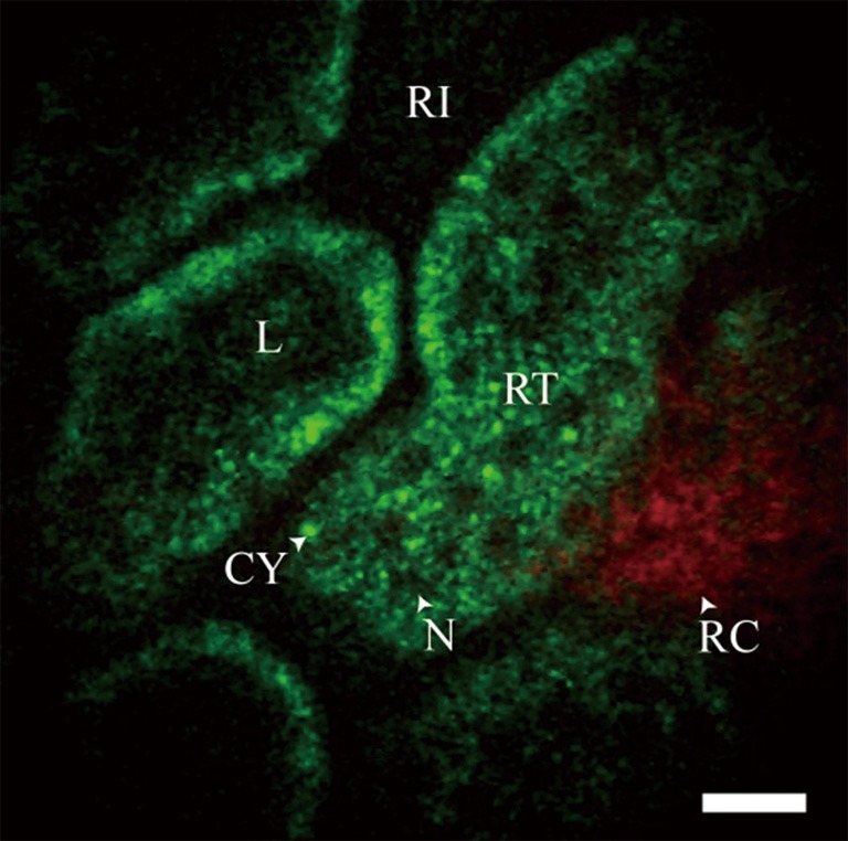
Figure 4.

In vivo image of unstained rat kidney with GRIN lens. The pseudo-color images show red SHG signal (<405 nm) and green intrinsic fluorescent emission (405-700 nm). Shown is the superficial renal cortex showing dark renal interstitium (RI), dark cellular nuclei (N) and bright intrinsic fluorescent cytoplasm (CY) that form the epithelial cells in the renal tubules (RT), SHG signal from the tough fibrous layer that forms the renal capsule (RC), and the dark blood filled lumen (L) inside the RTs. Scale bar is 20 µm (31).
