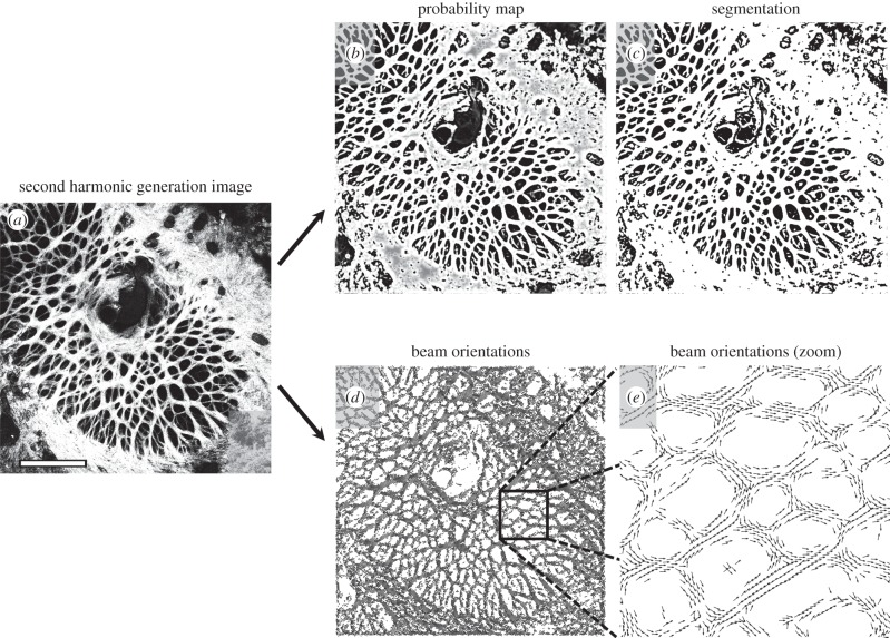
Figure 6.
Frangi's filter identifies beam location and orientation in confocal microscopy images. Similar to figure 5, we used Frangi's filter to enhance beam-like objects in this two-dimensional image (eye E). (a) The original image yields (b) a probability map that each voxel is a member of a beam, which can then be (c) binarized to segment the beams. Simultaneously, the eigenvectors of the Hessian matrix provide (d,e) information on local beam orientation. These vectors correspond to the smallest eigenvalue. In this visualization, only every 150th vector is shown due to the high resolution of the image. White scale bar, 1.0 mm.

