Figure 2.
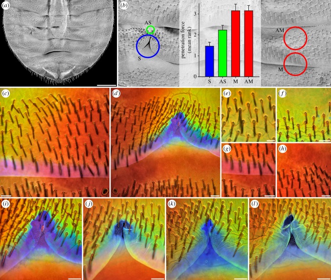
Material composition and properties of the ventral abdominal cuticle of C. lectularius females. (a) Abdomen overview (scanning electron micrograph). (b) Section of (a) indicating the locations of the spermalege (S), AS, M and AM, and penetration forces (mean ranks and standard errors, see Material and methods) determined for these four cuticle sites. (c–l) CLSM maximum intensity projections. (c,d) Autofluorescence composition of the cuticle in the left (c) and right (d) abdomen parts. The dominance of violet/blue autofluorescence (shown in blue) is restricted to the spermalege, clearly indicating that only at this site the cuticle contains large proportions of resilin. (e–h) Autofluorescence composition of the cuticle at the sites AS (e,f), M (g) and AM (h). The cuticle at M and AM consists mainly of sclerotized chitinous material, indicated by the dominance of red autofluorescence, while the presence of large proportions of green autofluorescence in the cuticle at AS indicates that the respective material consists mainly of weakly or non-sclerotized chitinous material. (i–l) Autofluorescence composition of the cuticle at the spermaleges of different one-week-old females, indicating variation of the extent of the resilin-dominated spermalege structures between females. Scale bars, (a) 500 µm, (c,d,f–l) 50 µm, (e) 25 µm.

