Figure 3.
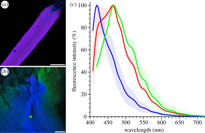
Properties of resilin autofluorescences. (a,b) Lambda-coded optical sections through a large part of the resilin-dominated structures of an elastic tendon of A. cyanea (a) and through a spermalege area of C. lectularius (b), showing autofluorescences excited by 355 nm laser light. In most of the tendon structures, violet resilin autofluorescence is dominant, while the terminal tendon parts exhibit mainly blue resilin autofluorescence. The spermalege is dominated by autofluorescence that is similar to the blue autofluorescence in (a), which indicates a resilin proportion similar to that in the terminal tendon parts. The surrounding cuticle exhibits green autofluorescence, indicating large proportions of other cuticle components such as chitin. The dots indicate locations where the fluorescence emission properties were analysed. (c) Emission spectra of the resilin autofluorescences exhibited by the elastic tendon (blue and red) and the spermalege (green); colours correspond to the dot colours in (a,b); mean values (lines, n = 5) and standard deviations (shaded areas). The spectral properties of the tendon's violet resilin autofluorescence are similar to those described for the ‘typical’ resilin autofluorescence [36,37], while the properties of the tendon's blue resilin autofluorescence are similar to those of the resilin autofluorescence present in the spermalege. Scale bars, (a) 100 µm, (b) 25 µm.

