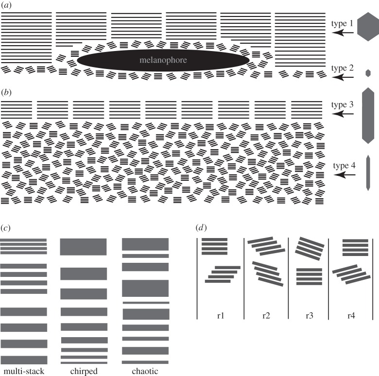
Figure 5.
The organization of the guanine platelets in the horizontal section of the lookdown's skin and models for broadband reflectance. (a) The arrangement of Type 1 and Type 2 guanine platelets and melanophores (me) in the dorsolateral skin. Long and short bars represent Type 1 and Type 2 guanine platelets, respectively. (b) The arrangement of Type 3 and Type 4 guanine platelets in the mid-lateral or ventrolateral skin. Long and short bars represent Type 3 and Type 4 guanine platelets, respectively. The morphologies of these guanine platelets are illustrated at the right side of the figure. (c) Three existing radial variation models for broadband reflectance by silvery fish: the multi-stack, chirped and chaotic models. (d) The lateral variation model for broadband reflectance. For simplicity, only two stacks of guanine platelets in four of many possible arrangements are illustrated (r1–r4). Variations in spacing and orientation among platelet stacks exist in these four different regions.

