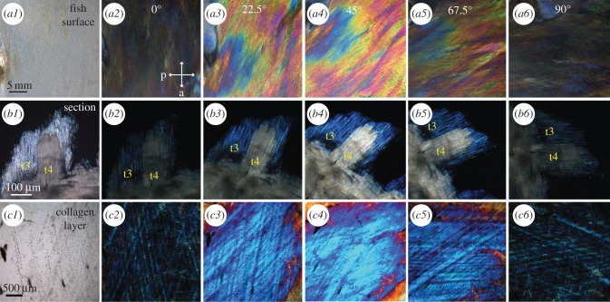
Figure 7.
Birefringence of the intact surface of the lookdown's skin (a2–a6), the skin section (b2–b6) and the collagen layer (c2–c6) visualized by cross-polarization microscopy. (a1) An image of the mid-lateral flank of a juvenile lookdown was taken with unpolarized light. The anterior–posterior axis of the fish was horizontal with the fish's head on the left. (a2–a6) Cross-polarization images of the same view as in (a1) were taken as the fish was rotated from the horizontal position (0°) clockwise by 22.5°, and 45°, 67.5°, or 90°, respectively. The maximum intensity was observed at 45° (a4). The orientations of the polarizer (p) and analyser (a) are indicated by the arrows in (a2). (b1) An image of a parasagittal section of the mid-lateral skin was taken with unpolarized light as the long axes of the platelets were aligned with the analyser. (b2–b6) Cross-polarization images of the same view as in (b1) were taken as the tissue section was rotated from its original position (0°) clockwise by 22.5°, and 45°, 67.5°, or 90°, respectively. The long axes of Type 3 (t3) and Type 4 (t4) platelets were in the same orientation. They both had minimum visibility when their long axes were in vertical or horizontal position, while the maximum visibility was reached as the section was rotated by 45o. Type 3 platelets appeared blue, while Type 4 platelets were multi-coloured at high magnification and appeared nearly white at low magnification (b4). (c1) An unpolarized-light image of the collagen layer from the mid-flank with the same orientation as in the fish (a1). (c2–c6) Cross-polarization images of the collagen layer as it was rotated by 22.5°, and 45°, 67.5°, or 90°, respectively. The maximum intensity was observed at 45°.

