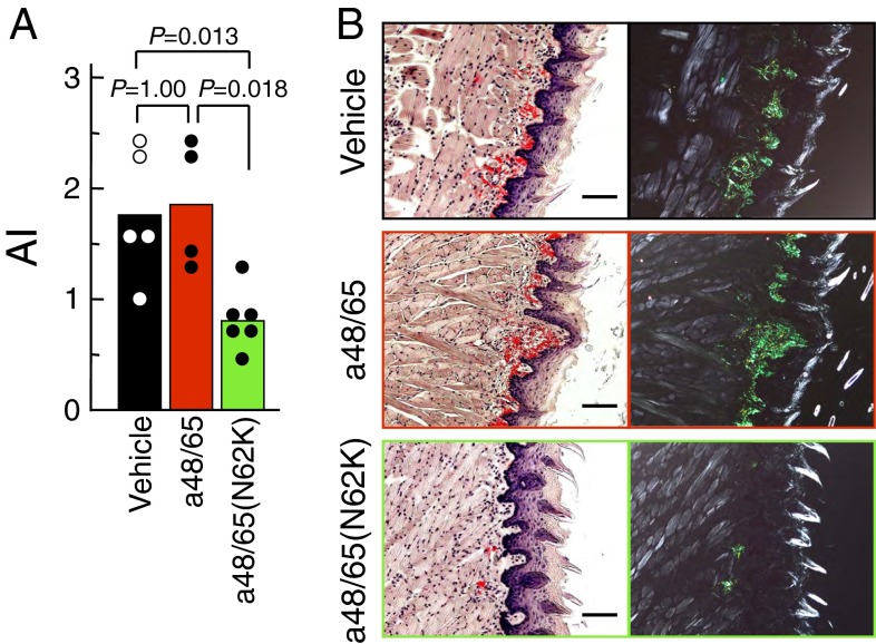
Fig. 8.
Suppression of amyloid deposits in vivo by treatment with a48/65(N62K). A single administration of 5 µg of AApoAII fibrils was given to 2-mo-old amyloidosis-susceptible mice using osmotic pumps implanted in their abdominal cavities. After 27 d, amyloid deposits were detected. (A) The AI in mice induced by AApoAII fibrils. Each bar and dot shows the mean of the group and individual grade of amyloid deposit. P value, Mann–Whitney U test. (B) LM images of amyloid deposits in the tongues by Congo red staining. (Left) Under normal light. (Right) Under polarized light. Grades of amyloid deposits in the tongues were 3, 3, and 2 in mice with vehicle, a48/65, and a48/65(N62K) pumps, respectively. (Scale bars: 100 µm.)

