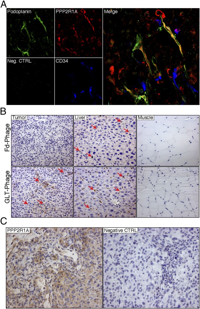
Fig. 4.
PPP2R1A is expressed on LyVs in vivo. (A) Confocal microscopy was performed on C8161 tumor frozen sections stained with PPP2R1A (red), Podoplanin (green), and CD34 (blue). Colocalization (yellow) of PPP2R1A and Podoplanin is depicted in the merged image. Neg. CTRL, negative control. (B) Immunohistochemical analysis of GLTFKSL phage homing. Positive staining (red arrows) is shown in the tumor of GLTFKSL-injected mice, whereas insertless phage staining in the tumor is negative. Muscle was the negative control organ. Liver is part of the reticuloendothelial system, and therefore, phage is present. Representative pictures are shown (magnification: 200×). (C) PPP2R1A expression in C8161 tumors. PPP2R1A is highly expressed in C8161 tumors as shown by immunohistochemistry. CTRL, control. (Magnification: 200×.)

