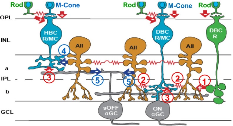
Fig. 1.
Schematic diagram of major synaptic connections in the ON and OFF α-ganglion pathways in the mouse retina. Green, rods and rod BCs; blue, M cones and mixed rod/M-cone BCs; orange, AIIACs; gray, αGCs; arrows, chemical synapses (red, glutamatergic; blue, glycinergic; +, sign-preserving; −, sign-inverting); zigzag (red), electrical synapses. a, sublamina a; b, sublamina b; GCL, ganglion cell layer; INL, inner nuclear layer; IPL, inner plexiform layer; OPL, outer plexiform layer; PRL, photoreceptor layer. Synapses directly relevant to this study are marked with numbers in circles: 1: DBCR→AIIAC glutamatergic; 2: DBCC↔AIIAC electrical; 3: DBCR/MC/HBCR/MC→ONαGC/sOFFαGC glutamatergic; 4: AIIAC→HBCR/MC glycinergic; and 5: AIIAC→sOFFαGC glycinergic.

