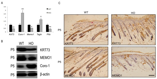
Fig. 3. Verification of proteomic analysis by qRT-PCR and immunohistochemistry analyses. (A) Relative mRNA expression of Krt73, Coro-1, Memo1, Phb and Tagln in the dorsal skin of wild-type and homozygous iRhom2Uncv littermates at P5. Each bar represents the mean ± SD (*P < 0.05, **P < 0.01 compared with wild-type mice). (B) The expression of KRT73, MEMO1 and Coro-1 was detected by western blot using specific antibodies. (C) Immunohistochemical staining of KRT73 and MEMO1 performed on skin sections of P5 wild-type and homozygous iRhom2Uncv mice. Scale bars=50 μm.

