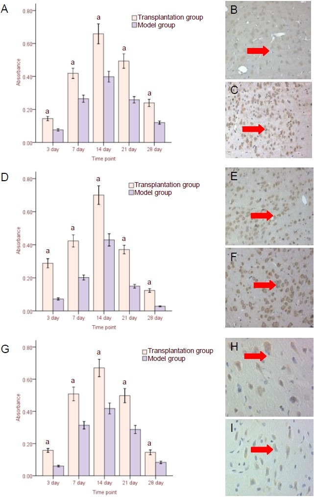
Figure 4.

Neurotrophic factor expression in surrounding injured brain tissues following human umbilical cord blood mesenchymal stem cell transplantation.
(A–C) Nerve growth factor (NGF) protein expression (immunohistochemistry, × 200); (D–F) brain-derived neurotrophic factor (BDNF) protein expression (immunohistochemistry, × 200); (G–I) BDNF mRNA expression (in situ hybridization, × 400).
(A, D, G) Results (absorbance) are expressed as mean ± SD from six rats in each group at each time point.
aP < 0.05, vs. model group (t-test was used to specify differences between two groups at the corresponding time points); (B, E, H) model group at 14 days after transplantation; (C, F, I) transplantation group at 14 days after transplantation. Arrows: NGF protein-, BDNF protein-, and BDNF mRNA-positive cells.
