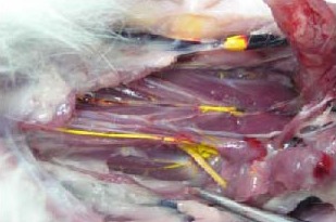
Figure 2.

Macroscopic view of Microfil distribution in cervical lymphatic vessels.
some Microfil fills and opacifies the head and cervical superficial lymphatic vessels. However, the majority of Microfil is observed in deep cervical lymphatic vessels along the posterior pharyngeal wall to the cervical lymphatic trunks and thoracic duct.
