
Figure 6.
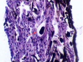
Histological observation of Microfil in nasal septum lymphatic vessels (hematoxylin-eosin staining, × 400).
Most lymphatic vessels within the olfactory submucosa comprise a single layer of endothelial cells, which is characteristic of lymphatic vessels. Dark-brown Microfil (red arrow) is visible in lymphatic vessels.