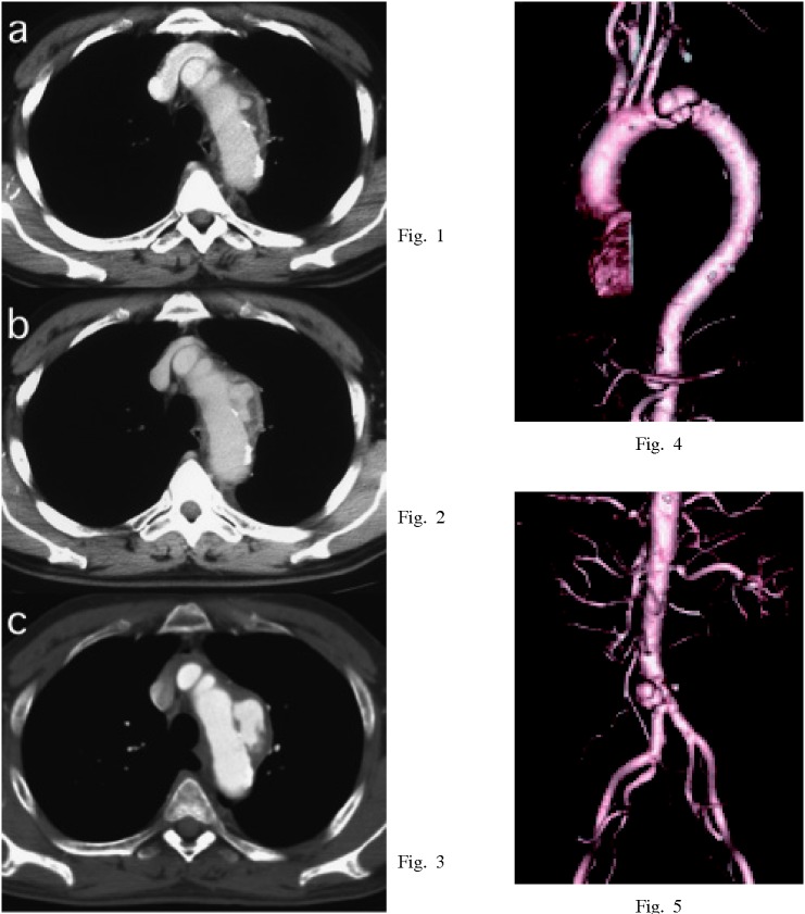
Fig. 1-5.
Enhanced chest CT findings of a 66-year-old male. Distal arch aorta revealed an ulcer-like protrusion with low density mass under thickened and enhanced adventitia at first (Fig.1). The ulcer-like protrusion expanded abruptly (Fig.2), and the diameter of the aneurysm increased from 40 mm to 70 mm within 10 days (Fig.3). The aneurysm developed as a result of invasion of bacteria into the aortic vessel wall (microbial arteritis with aneurysm). He also had an infrarenal abdominal aortic aneurysm. This aneurysm expanded from 30 mm to 40 mm in diameter within the same period (Fig.5).

