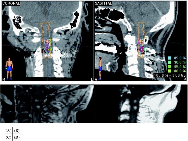
Fig. 2.
Case 1. Coronal view (A) and sagittal view (B) of CT with contrast enhancement during treatment plan of Novalis SRT for C1 and C2 intramedullary metastases on BrainSCAN (BrainLAB, Tokyo, Japan). Sagittal views of MRI before (C) and 4 months after SRT (D). The treated lesions were stable for 10 months after SRT until the patient died from primary lung adenocarcinoma.

