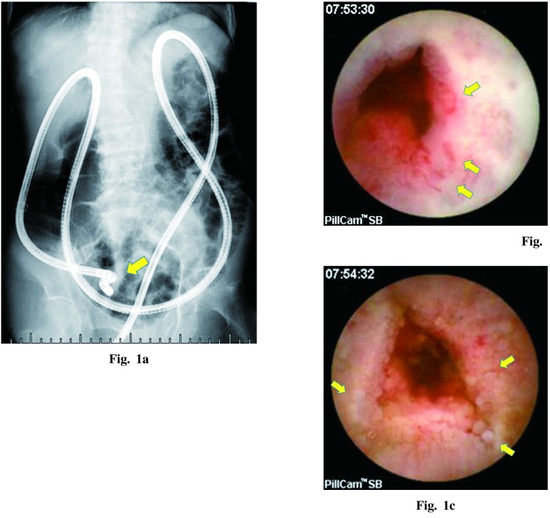
Fig. 1a Fluoroscopic image during retrograde DBE. The endoscope was crooked and was not inserted into the deeper ileum because of pelvic adhesion.
Fig. 1b Multiple angiectasias and fresh blood visualized with VCE.
Fig. 1c VCE indicated rough mucosa, which consisted of atrophic, irregular or scattered white villi.

