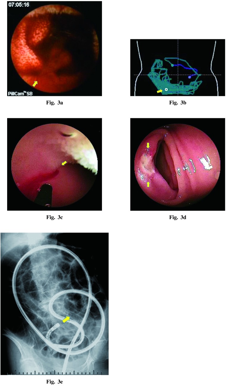
Fig. 3a VCE detected a spouting hemorrhage in the distal ileum.
Fig. 3b The localization map of the bleeding point in the VCE reading software.
Fig. 3c Retrograde DBE accessed the bleeding point that VCE indicated and attempted hemostasis using the heat coagulation device.
Fig. 3d The bleeding was stopped with endoscopic coagulation alone.
Fig. 3e Fluoroscopic image during retrograde DBE. The endoscope was smoothly inserted into the deeper ileum.

