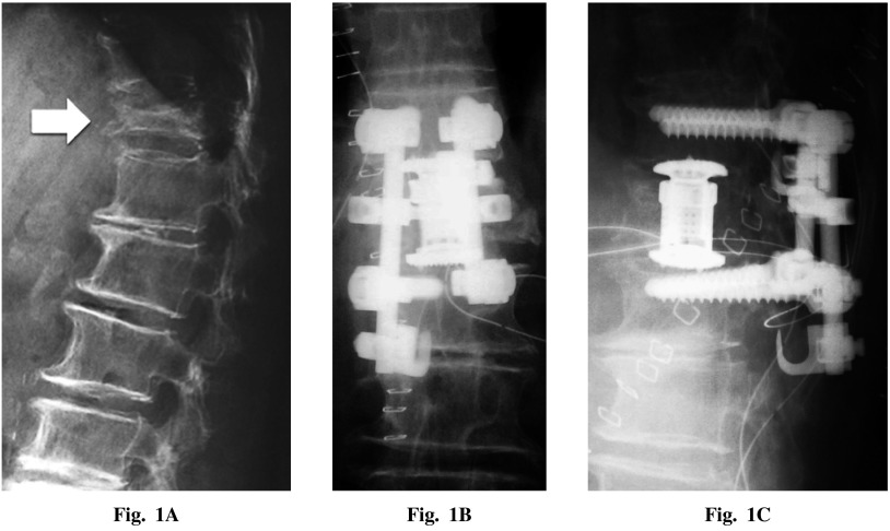
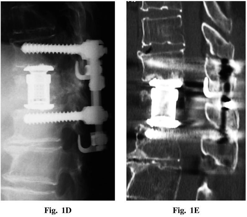
Fig. 1.
A: Preoperative lateral radiograph showed osteoporotic compression fracture at Th12 (white arrow). Postoperative posteroanterior (B) and lateral (C) radiographs. D: Postoperative lateral radiograph taken 1 year post surgery showing no loss of correction. E: Postoperative CT scan taken 1 year post surgery.


