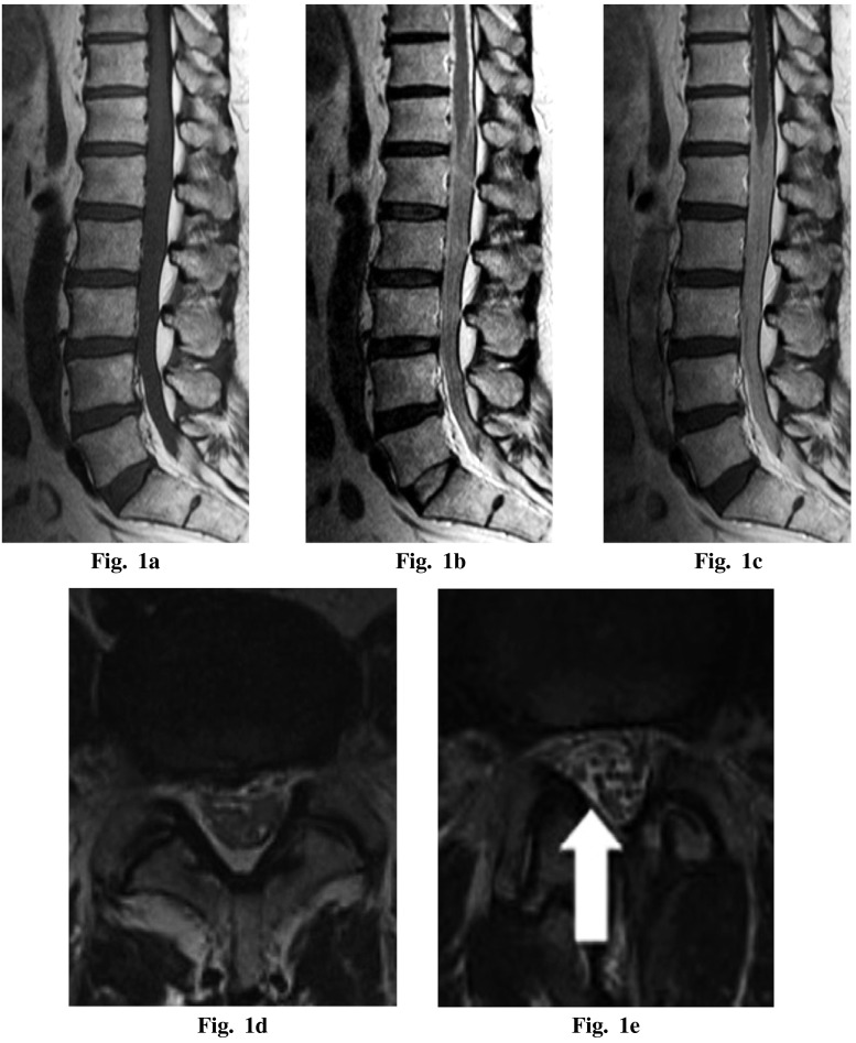
Fig. 1.
Magnetic resonance imaging — (a) preoperative T1-weighted sagittal image, (b) preoperative T2-weighted sagittal image, (c) preoperative T1-weighted sagittal image with gadolinium enhancement showing an intrathecal mass at the lesion and marked enhancement with gadolinium, (d) preoperative T2-weighted axial image at L4/5 level (e) postoperative T2-weighted axial image at L4/5 level. Swelling of the cauda equina was diminished (white arrow).

