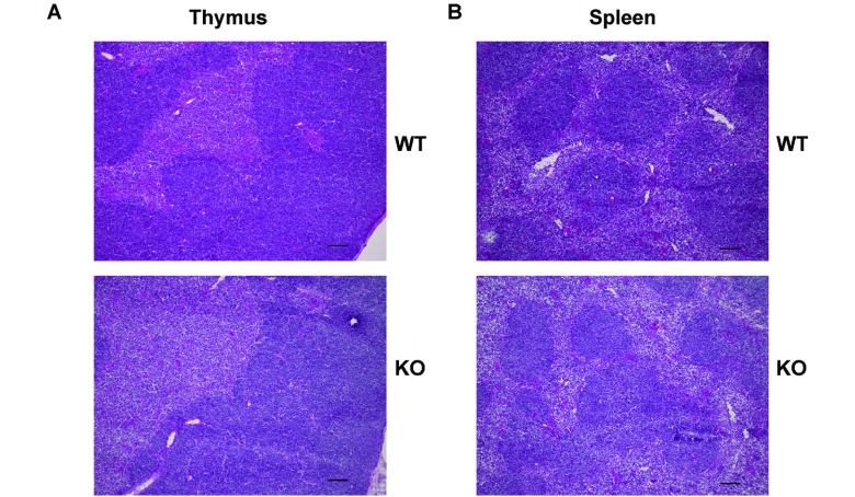
Fig. 2.
HE staining of the thymus and spleen.
A. HE staining of the thymus from the WT and KO mice (9 weeks old, female). Frozen sections were fixed in 4% PFA and stained with HE. The figures are representatives of two samples (from two mice in each group). Scale bars: 100 μm.
B. HE staining of the spleen from WT and KO mice (6 weeks old, female). Frozen sections were fixed in 4% PFA and stained with HE. The figures are representatives of two samples (from two mice in each group). Scale bars: 100 μm.

