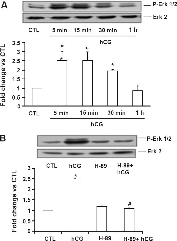
Fig. 4.

Activation of ERK1/2 in human granulosa cells. Day 4 granulosa cells were serum-starved, treated with hCG (10 IU/ml) alone for different time intervals (5 min, 15 min, 30 min, and 1 h; (A) or in the presence of H-89 (10 µM; 1 h pretreatment) for 15 min (B) and were lysed using RIPA buffer. The cell lysates were subjected to Western blot analysis to detect p-ERK1/2. The membranes were then stripped and reprobed for total ERK2. Lower panels represent densitometric scanning of the p-ERK1/2 signals normalized with ERK2 and expressed as fold change vs. CTL. The blots shown are representative of three independent experiments, and the results in the bar graphs are average and SE of three experiments. *, P < 0.05 vs. CTL; #, P < 0.05 vs. hCG. (Source: Ref. 27, Reproduced with permission).
