
Figure 1.
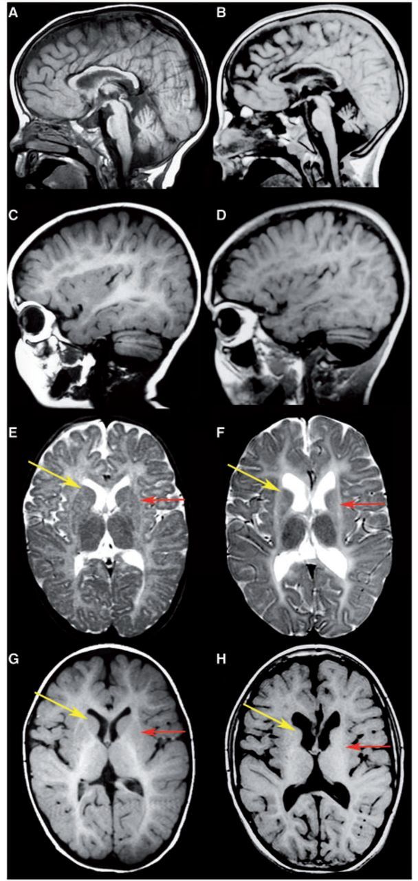
MRI findings in a typical H-ABC patient. Sagittal T1-weighted (A–D) and axial T2-weighted (E and F) and T1-weighted (G and H) images in Patient HA23 with the common c.745G > A mutation at the age of 1.5 years (A, C, E and G) and 13 years (B, D, F and H). The early MRI shows a mildy hyperintense white matter signal on T1-weighted (C and G) and T2-weighted (E) images, indicating a moderate lack of myelin. In the late MRIs, the white matter T1-signal is subtly reduced, indicating loss of some myelin (D and H). The cerebellar atrophy increases over time (A–D), especially of the vermis (A and B). At 1.5 years, a small putamen is present that has lost some of its normal grey matter signal (red arrows); the caudate nucleus is normal (yellow arrows in E and G). Note that at the age of 13 years, the putamen is no longer visible (red arrows) and that the caudate nucleus is slightly atrophic (yellow arrows in F and H). Over time, the lateral ventricles show a slight increase in size (E–H), indicating some loss of white matter volume.