
Figure 4.
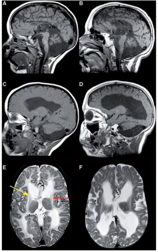
Patient with extensive MRI abnormalities. Sagittal T1-weighted (A–D) and axial T2-weighted (E and F) images of Patient HA51 with a c.1061G > A mutation at the age of 2 (A, C and E) and 7 years (B, D and F). The white matter has an intermediate to low signal on the T1-weighted images (C and D) and a high signal on the T2-weighted images (E and F), indicating a severe, almost complete, lack of myelin. There is serious loss of cerebral white matter volume from early on (C and E), which increases over time (D and F). The cerebellar atrophy is profound, involving both the vermis (A and B) and the hemispheres (C and D). By 2 years of age, the putamen (red arrow) is absent and the head of the caudate nucleus (yellow arrow) is entirely flat (E).