
Figure 3.
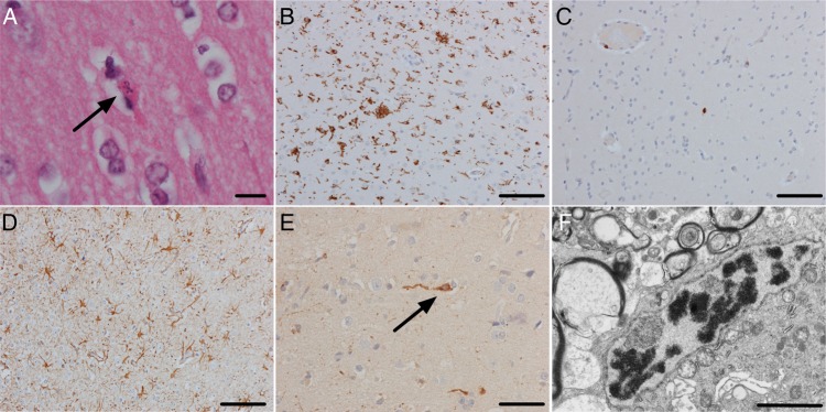
The brain biopsy showed extensive neuronal apoptosis in the cerebral cortex as demonstrated by pyknotic and karyorrhectic neurons (A, arrow) (hematoxylin and eosin [H&E], scale bar, 10 µm). There was a brisk microglial reaction demonstrated by CD68 immunohistochemistry with some nodules of microglia (B; scale bar, 100 µm). In contrast, there was no significant lymphocytic reaction (C, CD3 immunohistochemistry; scale bar, 100 µm). There was an extensive astrocytosis (D, glial fibrillary acidic protein [GFAP]; scale bar, 100 µm). Immunohistochemistry with an antibody against astrovirus showed extensive staining of cell bodies and processes in the neuropil (E; scale bar, 50 µm). Some of the positive cells had the morphology of pyramidal neurons (arrow). Electron microscopy showed a rare focus of crystalline material but no viral particles (F; scale bar, 2 µm).