Figure 3.
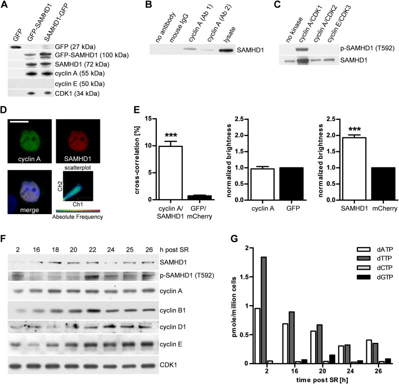
SAMHD1 is regulated by cyclin A in a cell cycle-specific manner. (A) Coimmunoprecipitation with N- or C-terminally green fluorescent protein (GFP)-tagged SAMHD1 expressed in HEK293T cells immobilised on GFP-Trap beads pulls down cyclin A and cyclin-dependent kinase 1 (CDK1), but not cyclin E. (B) Reverse coimmunoprecipitation with two different cyclin A antibodies (Ab 1, Ab 2) confirms interaction of SAMHD1 and cyclin A at the endogenous level. (C) In vitro kinase assay reveals phosphorylation of SAMHD1 at T592 by cyclin A/CDK1, but not by cyclin A/CDK2 or cyclin E/CDK3. (D) Cyclin A and SAMHD1 colocalise within the nucleus as shown by a high correlation of fluorescence signals in the scatter plot. Scale bar, 10 μm. (E) Fluorescence cross-correlation spectroscopy of living HeLa cells cotransfected with mCherry–SAMHD1 (SAMHD1) and GFP–cyclin A (cyclin A) shows a high cross-correlation, indicating formation of mobile SAMHD1–cyclin A complexes. Compared with monomeric GFP or mCherry, the average normalised brightness of GFP–cyclin A and mCherry–SAMHD1 is 1 or 2, respectively. At least 15 cells were measured per experiment. Data represent means±SEM from two independent experiments. ***p<0.001 by Student's t test. (F) HeLa cells synchronised at G0/G1 were lysed at the indicated time points after serum re-addition (SR) and immunoblotted with the indicated antibodies. (G) Intracellular deoxyribonucleoside triphosphate (dNTP) concentrations in HeLa cells treated as in (F).

