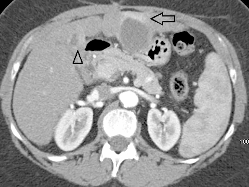
Fig. 3.

Arterial phase axial computed tomographic images performed 6 weeks after transarterial chemoembolization demonstrate near-complete resolution of the segment 3 lesion (arrow) and persistent peripheral nodular enhancement of the segment 4B lesion (arrowhead).
