Fig. 7.

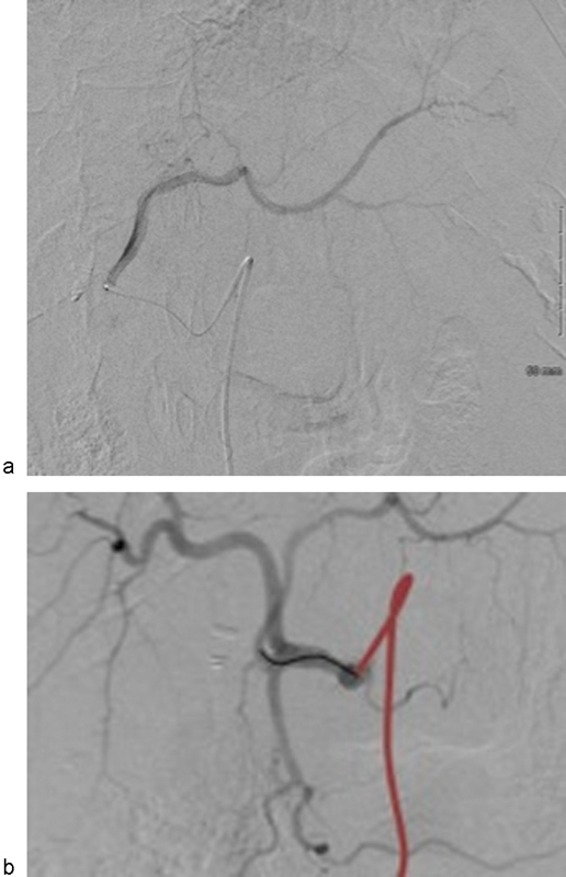
(a) Left hepatic arteriogram prior to TACE from the left hepatic artery. (b) Common hepatic arteriogram with red marking position of base catheter and black marking position of microcatheter during chemoembolization of the left hepatic lobe.

(a) Left hepatic arteriogram prior to TACE from the left hepatic artery. (b) Common hepatic arteriogram with red marking position of base catheter and black marking position of microcatheter during chemoembolization of the left hepatic lobe.