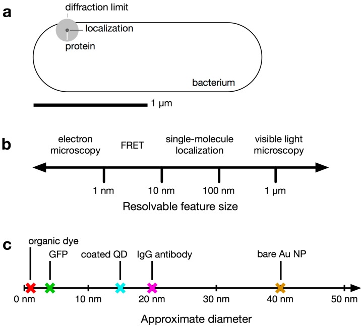
Figure 1.
Relevant size scales in subcellular imaging of bacteria. (a) Comparison of the size of a V. cholerae bacterial cell and the diffraction limit of light. A small fluorescent protein appears blurred to the diffraction limit (~200 nm, gray circle). Single-molecule imaging localizes the fluorophore with greater precision (~30 nm, black circle); (b) Resolvable size scales (log scale) using several common microscopy techniques; (c) Approximate diameters of an organic fluorophore, a fluorescent protein (GFP [8]), a coated quantum dot (QD; [9]), an immunoglobulin (IgG) antibody [10], and a 40-nm gold nanoparticle (NP).

